Back تصوير طبي بأشعة غاما Arabic Tomografia computada per emissió de fotó simple Catalan SPECT Danish Einzelphotonen-Emissionscomputertomographie German Tomografía computarizada de emisión monofotónica Spanish مقطعنگاری رایانهای تکفوتونی Persian Yksifotoniemissiotomografia Finnish Tomographie par émission monophotonique French טומוגרפיית פליטת פוטון בודד HE SPECT Hungarian
| Single-photon emission computed tomography | |
|---|---|
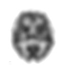
 A SPECT slice of the distribution of technetium exametazime within a patient's brain | |
| ICD-9-CM | 92.0-92.1 |
| MeSH | D01589 |
| OPS-301 code | 3-72 |


Single-photon emission computed tomography (SPECT, or less commonly, SPET) is a nuclear medicine tomographic imaging technique using gamma rays.[1] It is very similar to conventional nuclear medicine planar imaging using a gamma camera (that is, scintigraphy),[2] but is able to provide true 3D information. This information is typically presented as cross-sectional slices through the patient, but can be freely reformatted or manipulated as required.
The technique needs delivery of a gamma-emitting radioisotope (a radionuclide) into the patient, normally through injection into the bloodstream. On occasion, the radioisotope is a simple soluble dissolved ion, such as an isotope of gallium(III). Usually, however, a marker radioisotope is attached to a specific ligand to create a radioligand, whose properties bind it to certain types of tissues. This marriage allows the combination of ligand and radiopharmaceutical to be carried and bound to a place of interest in the body, where the ligand concentration is seen by a gamma camera.
- ^ SPECT at the U.S. National Library of Medicine Medical Subject Headings (MeSH)
- ^ Scuffham J W (2012). "A CdTe detector for hyperspectral SPECT imaging". Journal of Instrumentation. 7 (8). IOP Journal of Instrumentation: P08027. doi:10.1088/1748-0221/7/08/P08027. S2CID 250665467.
© MMXXIII Rich X Search. We shall prevail. All rights reserved. Rich X Search